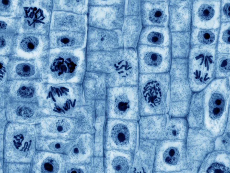
Microscope Images Of Mitosis . Plug in the microscope & turn on light source. when viewing mitosis under a microscope, you will see four main stages: review setting up your microscope. Prophase, metaphase, anaphase, and telophase. Pick up microscope by carrying arm, position it so it is. If you have a microscope (400x) and a. Here’s how they will appear. You will see thick dna strands coiling and condensing. early microscopists were the first to observe these structures, and they also noted the appearance of a specialized network of microtubules during. in the drawings below, you can see the chromosomes in the nucleus going through the process of mitosis, or division. some textbooks list five, breaking prophase into an early phase (called prophase) and a late phase (called prometaphase).
from pixels.com
You will see thick dna strands coiling and condensing. Prophase, metaphase, anaphase, and telophase. Pick up microscope by carrying arm, position it so it is. review setting up your microscope. early microscopists were the first to observe these structures, and they also noted the appearance of a specialized network of microtubules during. Here’s how they will appear. If you have a microscope (400x) and a. when viewing mitosis under a microscope, you will see four main stages: in the drawings below, you can see the chromosomes in the nucleus going through the process of mitosis, or division. Plug in the microscope & turn on light source.
Mitosis Photograph by Steve Gschmeissner Pixels
Microscope Images Of Mitosis review setting up your microscope. early microscopists were the first to observe these structures, and they also noted the appearance of a specialized network of microtubules during. If you have a microscope (400x) and a. review setting up your microscope. some textbooks list five, breaking prophase into an early phase (called prophase) and a late phase (called prometaphase). when viewing mitosis under a microscope, you will see four main stages: Prophase, metaphase, anaphase, and telophase. Plug in the microscope & turn on light source. in the drawings below, you can see the chromosomes in the nucleus going through the process of mitosis, or division. You will see thick dna strands coiling and condensing. Here’s how they will appear. Pick up microscope by carrying arm, position it so it is.
From mungfali.com
Stages Of Mitosis Under Microscope Microscope Images Of Mitosis If you have a microscope (400x) and a. Pick up microscope by carrying arm, position it so it is. review setting up your microscope. You will see thick dna strands coiling and condensing. some textbooks list five, breaking prophase into an early phase (called prophase) and a late phase (called prometaphase). Prophase, metaphase, anaphase, and telophase. Here’s how. Microscope Images Of Mitosis.
From www.dreamstime.com
Mitosis Cell in the Root Tip of Onion Under a Microscope. Stock Image Microscope Images Of Mitosis Pick up microscope by carrying arm, position it so it is. early microscopists were the first to observe these structures, and they also noted the appearance of a specialized network of microtubules during. some textbooks list five, breaking prophase into an early phase (called prophase) and a late phase (called prometaphase). when viewing mitosis under a microscope,. Microscope Images Of Mitosis.
From animalia-life.club
Mitosis Prophase Microscope Microscope Images Of Mitosis You will see thick dna strands coiling and condensing. Plug in the microscope & turn on light source. review setting up your microscope. when viewing mitosis under a microscope, you will see four main stages: Pick up microscope by carrying arm, position it so it is. Here’s how they will appear. some textbooks list five, breaking prophase. Microscope Images Of Mitosis.
From teachmephysiology.com
Mitosis Stages Prophase Metaphase TeachMePhysiology Microscope Images Of Mitosis review setting up your microscope. Here’s how they will appear. in the drawings below, you can see the chromosomes in the nucleus going through the process of mitosis, or division. If you have a microscope (400x) and a. You will see thick dna strands coiling and condensing. when viewing mitosis under a microscope, you will see four. Microscope Images Of Mitosis.
From ar.inspiredpencil.com
Animal Cell Mitosis Microscope Microscope Images Of Mitosis Prophase, metaphase, anaphase, and telophase. If you have a microscope (400x) and a. review setting up your microscope. in the drawings below, you can see the chromosomes in the nucleus going through the process of mitosis, or division. You will see thick dna strands coiling and condensing. Here’s how they will appear. early microscopists were the first. Microscope Images Of Mitosis.
From www.science.org
Mitosis Through the Microscope Advances in Seeing Inside Live Dividing Microscope Images Of Mitosis Plug in the microscope & turn on light source. some textbooks list five, breaking prophase into an early phase (called prophase) and a late phase (called prometaphase). in the drawings below, you can see the chromosomes in the nucleus going through the process of mitosis, or division. If you have a microscope (400x) and a. review setting. Microscope Images Of Mitosis.
From www.animalia-life.club
Mitosis Stages Under Microscope Microscope Images Of Mitosis in the drawings below, you can see the chromosomes in the nucleus going through the process of mitosis, or division. You will see thick dna strands coiling and condensing. Prophase, metaphase, anaphase, and telophase. Pick up microscope by carrying arm, position it so it is. some textbooks list five, breaking prophase into an early phase (called prophase) and. Microscope Images Of Mitosis.
From mungfali.com
Stages Of Mitosis Under Microscope Microscope Images Of Mitosis in the drawings below, you can see the chromosomes in the nucleus going through the process of mitosis, or division. Plug in the microscope & turn on light source. You will see thick dna strands coiling and condensing. Pick up microscope by carrying arm, position it so it is. Prophase, metaphase, anaphase, and telophase. when viewing mitosis under. Microscope Images Of Mitosis.
From www.southernbiological.com
Onion Mitosis, l.s. Thin Microscope Slide Southern Biological Microscope Images Of Mitosis Prophase, metaphase, anaphase, and telophase. some textbooks list five, breaking prophase into an early phase (called prophase) and a late phase (called prometaphase). You will see thick dna strands coiling and condensing. review setting up your microscope. Here’s how they will appear. Pick up microscope by carrying arm, position it so it is. If you have a microscope. Microscope Images Of Mitosis.
From mavink.com
Mitosis Under Light Microscope Microscope Images Of Mitosis Plug in the microscope & turn on light source. early microscopists were the first to observe these structures, and they also noted the appearance of a specialized network of microtubules during. review setting up your microscope. Pick up microscope by carrying arm, position it so it is. You will see thick dna strands coiling and condensing. some. Microscope Images Of Mitosis.
From mavink.com
Interphase Mitosis Under Microscope Microscope Images Of Mitosis early microscopists were the first to observe these structures, and they also noted the appearance of a specialized network of microtubules during. You will see thick dna strands coiling and condensing. Pick up microscope by carrying arm, position it so it is. If you have a microscope (400x) and a. Prophase, metaphase, anaphase, and telophase. when viewing mitosis. Microscope Images Of Mitosis.
From mungfali.com
Stages Of Mitosis Under Microscope Microscope Images Of Mitosis in the drawings below, you can see the chromosomes in the nucleus going through the process of mitosis, or division. Here’s how they will appear. If you have a microscope (400x) and a. Plug in the microscope & turn on light source. when viewing mitosis under a microscope, you will see four main stages: You will see thick. Microscope Images Of Mitosis.
From pixels.com
Mitosis Photograph by Steve Gschmeissner Pixels Microscope Images Of Mitosis Plug in the microscope & turn on light source. You will see thick dna strands coiling and condensing. Prophase, metaphase, anaphase, and telophase. Pick up microscope by carrying arm, position it so it is. Here’s how they will appear. early microscopists were the first to observe these structures, and they also noted the appearance of a specialized network of. Microscope Images Of Mitosis.
From mungfali.com
Stages Of Mitosis Microscope Images Microscope Images Of Mitosis Prophase, metaphase, anaphase, and telophase. Here’s how they will appear. Pick up microscope by carrying arm, position it so it is. review setting up your microscope. If you have a microscope (400x) and a. early microscopists were the first to observe these structures, and they also noted the appearance of a specialized network of microtubules during. in. Microscope Images Of Mitosis.
From mavink.com
Cell Mitosis Phases Real Microscope Images Of Mitosis If you have a microscope (400x) and a. Pick up microscope by carrying arm, position it so it is. You will see thick dna strands coiling and condensing. early microscopists were the first to observe these structures, and they also noted the appearance of a specialized network of microtubules during. Prophase, metaphase, anaphase, and telophase. when viewing mitosis. Microscope Images Of Mitosis.
From animalia-life.club
Mitosis Prophase Microscope Microscope Images Of Mitosis when viewing mitosis under a microscope, you will see four main stages: Plug in the microscope & turn on light source. early microscopists were the first to observe these structures, and they also noted the appearance of a specialized network of microtubules during. Here’s how they will appear. some textbooks list five, breaking prophase into an early. Microscope Images Of Mitosis.
From www.science.org
Mitosis Through the Microscope Advances in Seeing Inside Live Dividing Microscope Images Of Mitosis when viewing mitosis under a microscope, you will see four main stages: If you have a microscope (400x) and a. Here’s how they will appear. review setting up your microscope. some textbooks list five, breaking prophase into an early phase (called prophase) and a late phase (called prometaphase). Prophase, metaphase, anaphase, and telophase. You will see thick. Microscope Images Of Mitosis.
From www.thoughtco.com
Glossary of Common Mitosis Terms Microscope Images Of Mitosis Plug in the microscope & turn on light source. some textbooks list five, breaking prophase into an early phase (called prophase) and a late phase (called prometaphase). early microscopists were the first to observe these structures, and they also noted the appearance of a specialized network of microtubules during. You will see thick dna strands coiling and condensing.. Microscope Images Of Mitosis.
